
Overview
 Morton’s Neuroma is a common foot problem associated with pain, swelling and/or an inflammation of a nerve, usually at the ball-of-the-foot between the 3rd and 4th toes. Symptoms of this condition include sharp pain, burning, and even a lack of feeling in the affected area. Morton’s Neuroma may also cause numbness, tingling, or cramping in the forefoot.
Causes
Morton’s neuroma develops for several reasons. The primary reason is wearing narrow toe-box shoes, which compress the metatarsal heads. Certain anatomical factors also make nerve compression more likely with the narrow toe box shoes. In some people fibers, the medial and lateral plantar nerves converge close to the heads of the third and fourth metatarsals. This junction creates a larger nerve structure between the metatarsal heads making it more vulnerable to compression.
Symptoms
Patients with a Morton’s neuroma typically experience a sharp, shooting or burning pain, usually at the base of the forefoot or toes, which radiates into the two affected toes. Sometimes the pain may also radiate into the foot. The pain is often associated with the presence of pins and needles and numbness.
Diagnosis
A thorough subjective and objective examination from a physiotherapist is usually sufficient to diagnose a Morton’s neuroma. Investigations such as an X-ray, ultrasound, MRI, CT scan or bone scan may sometimes be used to assist with diagnosis, assess the severity of the injury and rule out other conditions.
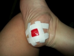
Non Surgical Treatment
Nonsurgical treatment is tried first. Your doctor may recommend any of the following. Padding and taping the toe area, shoe inserts, changes to footwear, for example wearing shoes with wider toe boxes or flat heels, Anti-inflammatory medicines taken by mouth or injected into the toe area, nerve blocking medicines injected into the toe area, other painkillers, physical therapy. Anti-inflammatories and painkillers are not recommended for long-term treatment. In some cases, surgery is needed to remove the thickened tissue and inflammed nerve. This helps relieve pain and improve foot function. Numbness after surgery is permanent..jpg)
Surgical Treatment
Surgery for Morton’s neuroma is usually a treatment of last resort. It may be recommended if you have severe pain in your foot or if non-surgical treatments haven’t worked. Surgery is usually carried out under local anaesthetic, on an outpatient basis, which means you won’t need to stay in hospital overnight. The operation can take up to 30 minutes. The surgeon will make a small incision, either on the top of your foot or on the sole. They may try to increase the space around the nerve (nerve decompression) by removing some of the surrounding tissue, or they may remove the nerve completely (nerve resection). If the nerve is removed, the area between your toes may be permanently numb. After the procedure you’ll need to wear a special protective shoe until the affected area has healed sufficiently to wear normal footwear. It can take up to four weeks to make a full recovery. Most people (about 75%) who have surgery to treat Morton’s neuroma have positive results and their painful symptoms are relieved.


 Overview
 Symptoms
 Overview
 Symptoms
 Prevention


